
OCT for monitoring the respiratory epithelium: OCT for monitoring the respiratory epithelium: Muco-ciliary clearance (MCC) is the self-clearing mechanism of the respiratory tract essential for the prevention of lung infections. When the MCC mechanism is defective, which can be associated with pulmonary diseases Cystic Fibrosis (CF) and Chronic Obstructive Pulmonary Disease (COPD), there are severe and devastating consequences for lung function. Mucus proteins create a matrix-like porous mesh that traps dangerous pathogens to protect the airways. When mucus becomes dehydrated due to disease, it collapses the MCC's cilia lining and pathogens are not cleared from the lungs. In order to monitor disease and determine treatment options, we must know more about the MCC system and the nanostructure of mucus in this dehydrated state.
Our laboratory has studied several markers that can be used to measure respiratory health as it relates to respiratory disease, in collaboration with the Cystic Fibrosis and Pulmonary Diseases Research and Treatment Center at UNC. We use OCT to explore the different health markers in air-liquid interface (ALI) cultures, which are human bronchial epithelial cells (HBECs) grown on a permeable membrane. These markers include the cilia beat frequency, mucus transporting rates (Oldenburg et al, 2012), and mucus weight percentage (Chhetri et al, 2014).
Using our PS-OCT system, we have demonstrated relative quantification of cilia beat frequencies (Oldenburg et al, 2012) at a single location on an HBEC. Our laboratory has recently constructed a parallel OCT system capable of capturing a B-mode image in a single snapshot. With this system, we have quantified cilia beat frequency over an entire cross-section of an HBEC (Barrick et al, 2016), as shown in Figure 1.

Fig. 1. Parallel OCT quantifies the median frequency (fm) of beating cilia on an HBEC across an entire cross section. The median frequency is related to the cilia beat frequency, which is a quantity relevant to medical diagnoses.
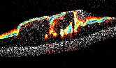
Most recently, our laboratory has employed DS-OCT to monitor clinically used mucus thinning treatments. DS-OCT captures the diffusion rate of gold nanorods (GNRs) as they move throughout mucus. DS-OCT is uniquely capable of resolving the diffusion rate of GNRs in space on short timescales. This capability allows us to monitor the changes in mucus during treatment therapies, as demonstrated in Figure 2 with isotonic and hypertonic saline (IS and HS) treatments. (Blackmon et al, 2017). We expect this novel technique of monitoring mucus weight percent in real-time will lead to better understanding of mucus thinning treatments, and eventually to an in vivo measure of treatment effectiveness for patients with respiratory disease.

Fig. 2. DS-OCT and corresponding HV-OCT (cross-polarized OCT intensity) monitor the changes in mucus weight percentage (wt%) via GNR diffusion during isotonic (~1% salt) and hypertonic (~5% salt) saline treatments. DS-OCT reveals high wt% mucus (red) that was lifted to the surface of the sample upon treatment and the decrease in wt% over time (red to orange/yellow). Differences in the two treatments, including a decrease in layer height with IS, and increase in layer height and mucus mixing, are revealed with DS-OCT. Layer features, and evidence of cell secretion (low HH-OCT intensity) is revealed with HV-OCT.
intro page - research - publications - people - open positions
UNC Physics & Astronomy - Biomedical Research Imaging Center - UNC Home