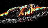

Parallel line-field spectral-domain OCT system: OCT is a noninvasive, high-resolution biomedical imaging modality which produces 2D cross-sectional images. Our first parallel OCT system (Barrick et al, 2016) provides images that span ~4 mm along the surface of tissue and ~ 0.5 mm deep into the tissue with an ultrahigh resolution of ~17 µm transversally and ~2 µm in depth. Put into a biological context, OCT is great for imaging layered soft tissue such as skin, fingernail beds, airways, and eyes. Having layered or otherwise inhomogeneous samples is key because OCT works by detecting a difference in the refractive index of the sample, so homogeneous samples typically don’t produce images as interesting as those of layered samples.
Parallel OCT is very similar to traditional, flying-spot OCT with respect to theory and the mechanism by which we are able to record images. The main difference between parallel OCT and flying-spot OCT is that the imaging beam is focused into a line at the sample rather than a small spot which has to be mechanically scanned across the surface of the sample. Essentially it is a faster way to record OCT images. Speed is important in biomedical imaging because living, moving cells will exhibit motion artefacts if you can’t scan faster than the cells are naturally moving.
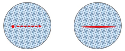
Fig. 1. Illustration of the main difference between flying-spot OCT (circular spot scanned across tissue surface) and parallel OCT (light focused into a line on the surface of the sample).
Parallel OCT is relatively new in the OCT community, but it is of increasing interest. The Oldenburg lab has only recently branched over into parallel OCT, and we currently have a working prototype parallel OCT system. As shown in the photo below, the system is a free-space optical system composed of many optical elements (lenses, mirrors, beam splitter) and a spectrometer.
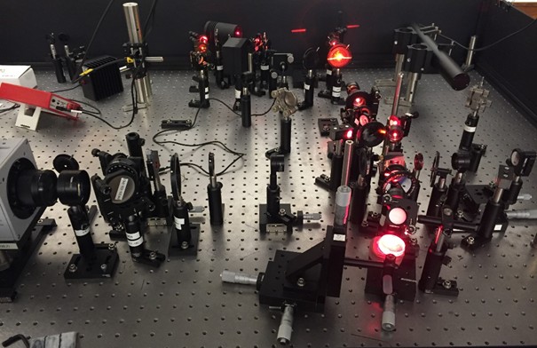
Fig. 2. Photograph of the Parallel OCT system.
Because the imaging beam is spread into a line rather than being focused into a small spot, parallel OCT requires a higher power source than traditional flying-spot systems in order to achieve similar sensitivities. We combine a high-power, supercontinuum light source from NKT Photonics with a free-space Michelson interferometer. We then use a cylindrical lens to focus the beam only in one transverse plane. This produces the line illumination unique to parallel OCT.
intro page - research - publications - people - open positions
UNC Physics & Astronomy - Biomedical Research Imaging Center - UNC Home