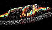

Imaging Nanoporosity using Diffusive Probes and OCT: Our body’s tissue is made up of proteins, primarily collagen, that are linked together to form a spongy mesh. The spacing between the proteins, i.e. the mesh pore sizes, can be used to study and monitor diseases such as breast cancer (Chhetri et al, 2014; Blackmon et al, 2016) and cystic fibrosis (Chhetri et al, 2014; Blackmon et al, 2017). Our laboratory has developed a method to measure these nanoscale pore sizes using gold nanoparticles. Nanoparticles strongly scatter light at the wavelengths used with our system due to a phenomenon is known as plasmon resonance. Instead of employing commonly used spherical particles, we use rod-shaped nanoparticles (GNRs) that have customized longitudinal plasmon resonance tuned to the centered of our OCT system’s wavelength. This makes them bright contrast agents against native tissue (Oldenburg et al, 2016) and we can track the GNRs natural Brownian motion in an environment as translational or rotational diffusion. In liquids with a high viscosity the rate at which these GNRs diffuse slows (Chhetri et al, 2011), while in tissue, the rate of diffusion is governed by the mesh pore sizes.
Spectral-domain OCT is a rapid imaging technique that allows us to collect depth-resolved 1-dimensional intensities in a single scan. Polarization-Sensitive Spectral-Domain OCT is well-suited to monitor these diffusion rates because the depth resolution and high-speed imaging rates capture the rapid diffusion of GNRs within small volumes. The polarization sensitivity of our OCT system allows us to disentangle both translational and rotational GNR diffusion and gives us an output cross-polarized OCT signal that is unique to the GNRs, giving us the ability to discriminate between them and tissue.
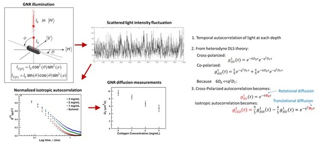
Figure 1.Measuring Gold Nanorod (GNR) diffusion rates in varying concentrations of collagen.
In order to measure this heterogenous nanoporosity, we have developed diffusion-sensitive optical coherence tomography (DS-OCT). For cystic fibrosis, this means we can monitor changes in the mucus mesh during mucus thinning treatments over time. For studying breast cancer, we obtain a 2D cross-sectional view of the extracellular matrix nanoporosity in space. The construction of DS-OCT images is illustrated below in Figure 2.
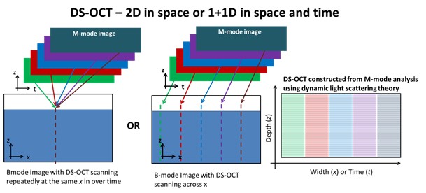
Figure 2. Illustration of how DS-OCT images are constructed from M-mode OCT scans for both resolution in 2D space and resolution in depth vs. time.
intro page - research - publications - people - open positions
UNC Physics & Astronomy - Biomedical Research Imaging Center - UNC Home