
Ultrahigh-resolution, polarization-sensitive, spectral-domain OCT system: The custom-designed spectral domain PS-OCT system shown in Figure 1 uses low coherence interferometry to collect cross-sectional images up to ~2 mm beneath tissue surfaces. The light source for UHR-PS-OCT is a low-coherence 800 nm, 120 nm bandwidth, Ti:Sapphire laser. Because this imaging technique uses LCI, we are able to depth resolve the features beneath the tissue surface using a technique called coherence gating. The wavelength of the near-infrared light source falls into a “biological optical imaging window” since it is minimally absorbed by the different components of tissue (water, melanin, hemoglobin, etc.), allowing for greater tissue penetration depths. The system resolution is ~3 × 10 µm axial × transverse (Chhetri et al, 2012), which is significantly greater than many conventionally used OCT systems, whose resolutions are ~10’s of microns.
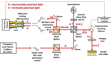
Figure 1: UHR-PS-OCT schematic, showing the propagation of horizontally and vertically polarized light through a Michelson interferometer to the custom built spectrometer.
This ultra-high resolution allows us to study micron-scale features in tissue, which is valuable for investigating respiratory disease, breast cancer, and dentistry. Since we are using an interferometric based system, we can monitor changes in the phase of light recorded, which gives us nano-scale sensitivity to movements within tissue, and has been used to develop new techniques to monitor the movement of nanoparticles introduced into tissue, like magnetomotive OCT to detect magneto-receptors, and diffusion-sensitive OCT to measure the nanoscale porosity of mucus or extracellular matrix. We obtain polarization sensitivity by isolating one polarization state at the input of our imaging system. This gives us the ability to discriminate between scatterers with different optical properties, like gold nanorods used with DS-OCT. This OCT system was custom built by our laboratory, shown in Figure 2, and therefore fully customizable to meet ever-changing needs.
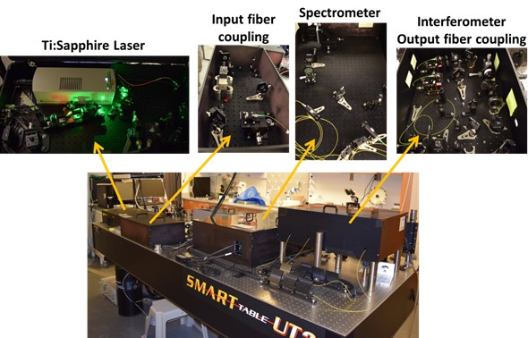
Figure 2: Snapshots of our custom built UHR-PS-OCT system.
intro page - research - publications - people - open positions
UNC Physics & Astronomy - Biomedical Research Imaging Center - UNC Home