
Polarization-Sensitive OCT for Assessing Tooth Restorations: It is estimated that general dentists spend 50-60% of their time replacing restorations that fail mainly due to fractures and secondary (recurrent) caries. These failures are exacerbated by micron-scale defects and gaps that can grow under fatigue from chewing and/or demineralization processes, ultimately resulting in restoration fracture and/or secondary caries formation. Optical coherence tomography (OCT) is rapidly growing as a method for assessing defects within restorative materials because it is nondestructive, high speed, translatable to clinical imaging, and of sufficient resolution to detect cracks. Yet, typical OCT systems employed for restorative studies are limited to ~10 micon resolution, which diminishes the ability to detect micron-scale defects. Our laboratory employs an ultrahigh resolution, polarization-sensitive, (UHR-PS-) OCT system (2 micron axial resolution in teeth) to study defects in different types of dental restorations before and after mechanical stress.
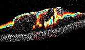
UHR-PS-OCT is able to clearly differentiate the enamel, resin-based restoration, and dentin in the tooth via cross-polarized contrast in Figure 1 (left column arrows). Images after mechanical stress reveal micron-scale voids (low scattering, right column arrows) that would be undetectable with conventional low-resolution OCT systems.

Fig. 1. UHR-PS-OCT before and after mechanical stress. PS-OCT images clearly differentiate the boundaries between restorative material and the native tooth (orange arrows in “Before” column) and the defects in the restoration after mechanical stress (orange arrows in “After” column). In these images, "HH" and "HV" correspond to co- and cross-polarized images obtained with horizontal (H) polarization incident upon the sample.
UHR-PS-OCT is also useful in detecting microcracks in the restoration, such as those shown in Figure 2 in a specimen after being polished with a fluted-carbide bur. This work was recently published by Vasconcellos et al, 2016.

Fig. 2. OCT imaging revealed voids and cracks throughout a restoration after mechanical stress (polishing with fluted-carbide bur), which were validated using microscopy. The ultra-high resolution provided with our OCT system may reveal defects that would be missed with conventionally used dental OCT systems.
Future studies will focus on longitudinal monitoring of restoration integrity for different materials and mechanical stress tests. Additionally, we are developing a user-friendly computer application for dental researchers and clinicians to assess 3D OCT data, similar to our segmentation tools for other applications, including luminal cross-sections (Wijesundara et al, 2014), organoids in tissue culture (Oldenburg et al, 2015), and retinal cysts (Wilkins et al, 2012). This application will include auto-segmentation and 3D visualization of restorative materials and their defects, as demonstrated in Figure 3, which will speed the translation of the findings of this research into clinical practice.

Fig. 3. The 3D reconstruction of dental restoration reveals marginal gaps along the edge of the restoration, and can be used to give dentists visualization of the restoration and potential defects.
intro page - research - publications - people - open positions
UNC Physics & Astronomy - Biomedical Research Imaging Center - UNC Home