
Detecting Pre-Occlusive Thrombosis via MMUS of SPIO-labeled Platelets: Blood clots, or "thrombi," are a leading cause of heart attacks and strokes, so detecting clots quickly and reliably is highly important. Currently, there are several detection techniques for thrombi, but they all have drawbacks. For example, Doppler ultrasound can only image anomalies in blood flow around clots – not the clots themselves, compression ultrasound cannot provide quantitative data, while x-rays and CT scans are expensive and give the patient high doses of radiation. The magnetomotive ultrasound (MMUS) system we are developing would use only a contrast agent, and a cost-effective, non-invasive, non-irradiating ultrasound exam to image blood clots, even in the early stages while they are still small.
Blood platelets which can be loaded with super-paramagnetic iron oxide particles (SPIOs) are a promising new contrast agent for detecting thrombosis via magnetomotive imaging. Specifically, thrombi form in response to arterial damage, when large quantities of platelets congregate at the damage site and form a stiff plug to prevent further bleeding. We plan to infuse blood platelets with SPIOs, producing a large quantity of “SPIO-labeled” platelets. These platelets will be inserted into the blood stream, where they will mix with the regular un-labeled platelets. Now, when a clot forms, a fraction of its platelets will be magnetically labelled. Since platelets congregate at high concentrations in thrombi, clots will contain high levels of magnetic particles, as shown in Figure 1, while other parts of the body will not.
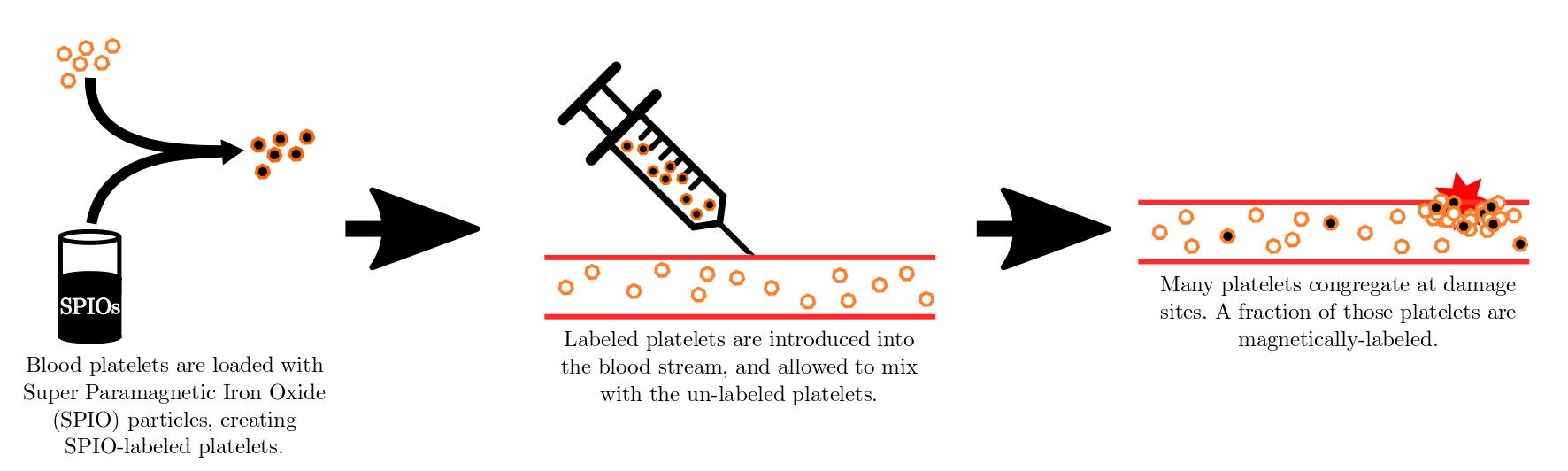
Fig. 1. This flow chart shows how magnetic particles are loaded into platelets, and added to the regular platelets in the blood stream. Any clots that form will now have high levels of magnetic particles, allowing them to be seen via MMUS.
The MMUS system we developed images an area of tissue using ultrasound, and then determines which areas of that image contain magnetic particles. The system uses two electromagnets, driven to create a sinusoidally-varying magnetic force, which causes a proportional sinusoidal displacement in the SPIO-labeled platelets. We then use frequency and phase-locking algorithms to determine which portions of the image exhibit this characteristic motion. These are the clots.
Past experiments in our lab have already shown that this method of imaging using SPIO-labeled platelets as contrast agents is possible. Oldenburg et al, 2010 showed that Optical Coherence Tomography (OCT) – an imaging method analogous to Ultrasound, but using optics rather than acoustics – showed that SPIO-platelets specifically adhered to a thrombus formed on a damaged vascular endothelium and provided contrast, compared to no contrast from that on un-damaged endothelium. Importantly, we have recently demonstrated that MMUS can sense simulated thrombus in the presence of pulsative flow (Levy et al, 2018). An MMUS image of such a model SPIO-labelled clot beneath a model thin plastic artery is shown in Figure 2.
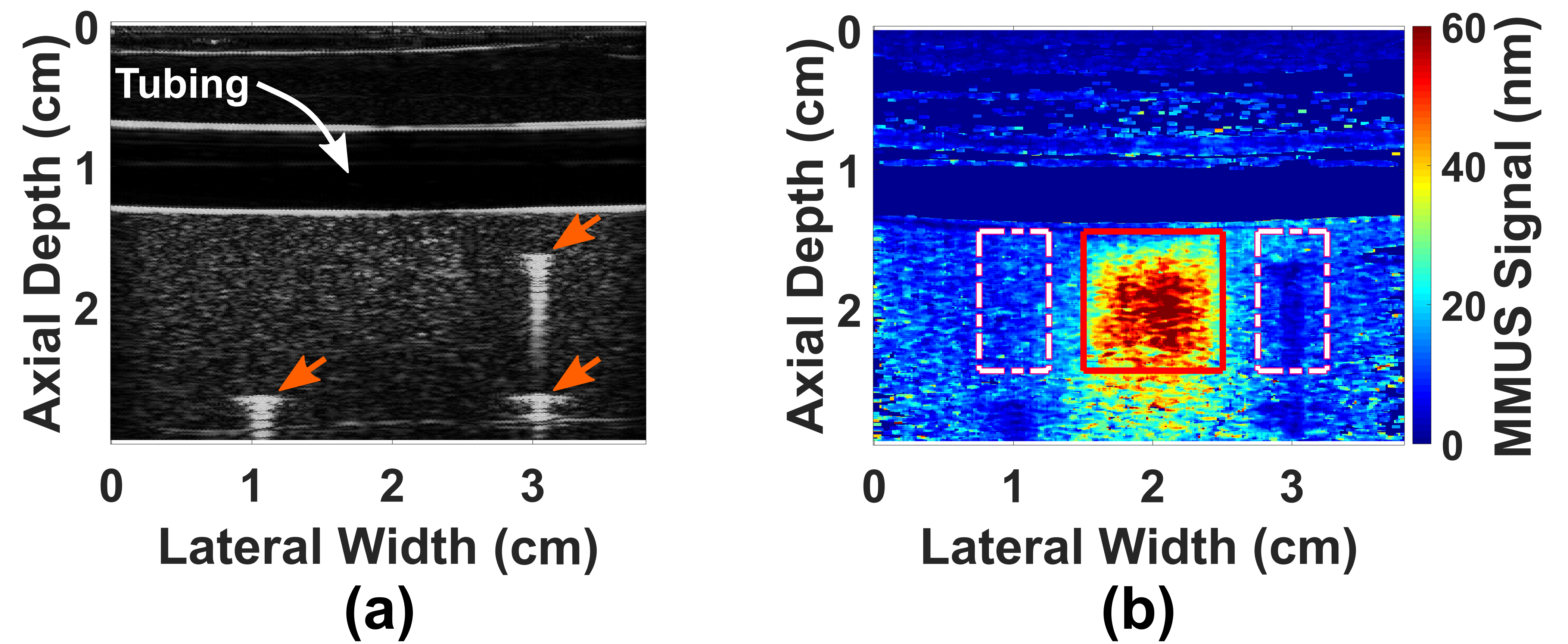
Fig. 2. A thin plastic tube embedded in gelatin models a blood vessel passing through human tissue. Water pumps though the tube (simulating blood flow), and a cubical gelatin model thrombus containing iron oxide particles is placed below the vessel. Figure (a) shows what is seen with conventional ("B-mode") ultrasound, and figure (b) shows what is seen with MMUS. The model thrombus is only visible in the MMUS image, and it appears as a red and yellow square beneath the tube. The true location of the model thrombus (shown by the thin red outline) can be seen to align well with the detected location. More on these images may be found in our recent publication: (Levy et al, 2018).
intro page - research - publications - people - open positions
UNC Physics & Astronomy - Biomedical Research Imaging Center - UNC Home